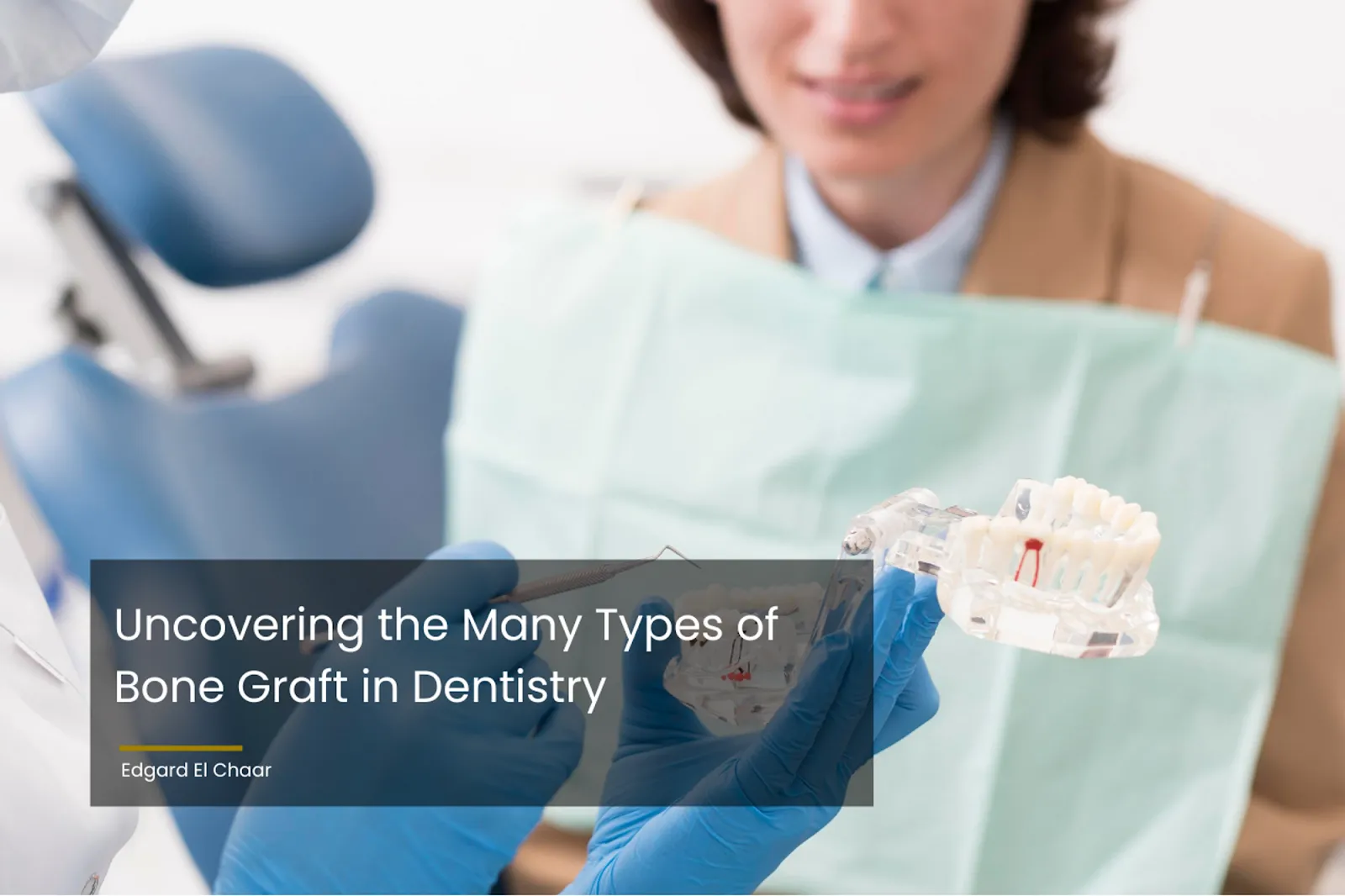
Uncovering the Many Types of Bone Graft in Dentistry

Laser Treatment for Gum Disease: Definition & Benefits
06/30/2023
Dry Socket with Bone Graft: Causes, Prevention & Treatment
07/02/2023The landscape of dentistry is constantly evolving, and bone grafting represents a significant advancement in this field. This article explores the various types of bone graft in dentistry and illustrates how our private practice of Edgard El Chaar consistently integrates these technological advancements to provide top-notch dental services.
What is Bone Grafting?
Bone grafting is a surgical procedure that enhances the quantity or quality of bone around the teeth. It’s particularly crucial for individuals who have experienced bone loss due to gum disease, tooth extraction, or trauma.
Why Do You Need Bone Grafting?
Bone grafting is a critical procedure in dentistry that is often employed as a precursor to other more intricate dental operations, such as the placement of dental implants. This procedure is particularly significant in cases where there is an insufficient natural bone in the area where the dental implant is to be placed.
This is where bone grafting steps in, providing a viable solution to rectify this issue. During the bone grafting procedure, the dentist will augment the affected area with the patient’s own bone, a donated bone, a synthetic bone substitute, or an animal bone. This material assists the body in rebuilding its own natural bone.
Over time, the grafted bone material will integrate with the patient’s natural bone, creating a stronger, more substantial foundation for the dental implant. This, in turn, can significantly improve the chances of a successful and long-lasting dental implant surgery.
⇒ Maybe you’ll be interested in: Can You Get Dental Implants With Gum Disease?
The Different Types of Bone Graft in Dentistry
In the dental field, four primary types of dental bone grafts are utilized, each with its unique characteristics and applications.
- Autografts
Autografts are bone grafts sourced from the patient’s body. This type of graft has a high success rate because it eliminates the risk of the body rejecting the graft material.
- Allografts
Allografts involve bone taken from a human donor. These grafts undergo rigorous testing to ensure safety and are beneficial when a significant amount of bone is needed.
- Xenografts
Xenografts are derived from non-human species, usually bovine sources. This type of graft is beneficial when the patient’s health prevents the use of an autograft.
- Alloplasts
Alloplasts are synthetic bone grafts, usually made from bioactive glass or other ceramics. They are advantageous for patients who prefer not to use natural bone for their graft.
Are There Risks of Bone Grafting?
As with any surgical intervention, bone grafting procedures carry certain potential risks and complications, despite being generally deemed safe and successful. These include:
- Infection: A common risk in surgeries, infection is preventable through sterile protocols and postoperative care, and is usually treatable with antibiotics if it occurs.
- Nerve Damage: This is a possible risk, especially in lower jaw surgeries, leading to temporary or sometimes permanent numbness in the lower lip, chin, and teeth.
- Graft Failure: A rare occurrence where the grafted bone doesn’t integrate with the natural bone. The risk increases in smokers or those with certain medical conditions.
- Sinus Problems: Relevant in upper back jaw surgeries, where graft material could enter sinus cavities, potentially causing complications.
Despite these risks, advancements in dental technology and the skill of a competent dental practitioner can significantly reduce them. Preoperative assessments, rigorous sterilization protocols, and patient selection help ensure successful outcomes.
How Long Does it Take for a Bone Graft to Heal?
The healing period following a bone graft can vary greatly, depending on individual health factors, lifestyle, and the type of graft used. In general, it takes several months for the grafted bone to fully integrate with the natural bone, a process known as osseointegration.
Post-procedure, patients need to follow a special care regimen to prevent infection and promote healing, including prescribed antibiotics, gentle oral hygiene, and dietary adjustments. For more complex grafting procedures, healing can extend to six to nine months, requiring regular dentist follow-ups to monitor progress. Smoking can delay healing and impede successful bone growth.
Therefore, the healing timeline is highly individualized and patients are encouraged to closely follow care instructions and maintain open communication with their dentist to ensure a successful grafting procedure.
Conclusion
At Edgard El Chaar‘s private practice, we specialized in allograft, to offer comprehensive care that meets the unique needs of each patient. Call us today to experience the cutting-edge techniques we employ and the excellent dental service that’s unparalleled by any others.
-
- Call Us: 212.685.5133 or 212.772.6900
-
- Contact Us by Submitting This Contact Form
Source:
Jallad, W. A. (2014, September 2). Restoration of Missing Upper Anterior Teeth using Dental Implant Simultaneous with Bone Grafting- A Case Report. Journal of Dentistry & Oral Health, 1–8. https://doi.org/10.17303/jdoh.2013.303
Eppley, B. L. (1996, January). Alveolar cleft bone grafting (Part I): Primary bone grafting. Journal of Oral and Maxillofacial Surgery, 54(1), 74–82. https://doi.org/10.1016/s0278-2391(96)90310-9
Ochs, M. W. (1996, January). Alveolar cleft bone grafting (Part II): Secondary bone grafting. Journal of Oral and Maxillofacial Surgery, 54(1), 83–88. https://doi.org/10.1016/s0278-2391(96)90311-0
Berger, J. S. (2010, September). The Use of Bone Morphogenic Protein for Bone Grafting Alveolar Defects. Oral Surgery, Oral Medicine, Oral Pathology, Oral Radiology, and Endodontology, 110(3), 312. https://doi.org/10.1016/j.tripleo.2010.05.016
